


Implant positions
Intraoral photogrammetry


Shining 3D Aoralscan Elite Intraoral Dental 3D Scanner

Available on Click & Collect · Pay by card or cash at pickup, or choose bank transfer at checkout How does it work?
Intraoral photogrammetry
Intraoral scanning
Coded scanbody workflow
| Scan Field of Single Frame | IPG scanner tip: 19mm × 14mm Standard scanner tip: 16 mm × 12 mm Mini scanner tip: 12 mm × 9 mm |
|---|---|
| Scan Depth | 22 mm from exit surface of tip |
| Scan Principle | Non-contact scanner with structured light and Intraoral photogrammetry |
| Dimension (L × W × H) | 245 mm x 30 mm x 26 mm |
| Weight | 124 g (without cables) |
| Output | STL, OBJ, PLY |
| Connection Port | Type-C |
Intraoral scanning and intraoral photogrammetry in one device.

Intraoral photogrammetry captures implant positions using the compatible coded scanbody workflow.

Switch to intraoral scanning for conventional restorative cases and soft-tissue capture.

The handpiece design supports positioning and access throughout the scan.
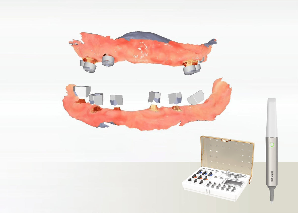
The Elite system combines normal intraoral scanning with intraoral photogrammetry for full-arch implant workflows. Select the appropriate mode and accessories for restorations, orthodontic records or All-on-X data capture.
The coded scanbody and Cap Scanbody workflow links implant reference positions to the surrounding soft-tissue scan. Compatible libraries convert the references into CAD data for downstream prosthetic design.
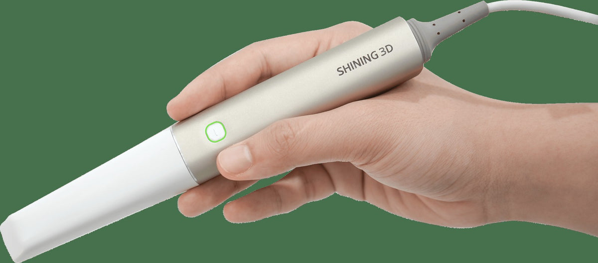
The larger IPG tip is designed for the photogrammetry and edentulous workflow. Standard intraoral scanning remains available for everyday cases, with the scanning strategy chosen by the clinician.
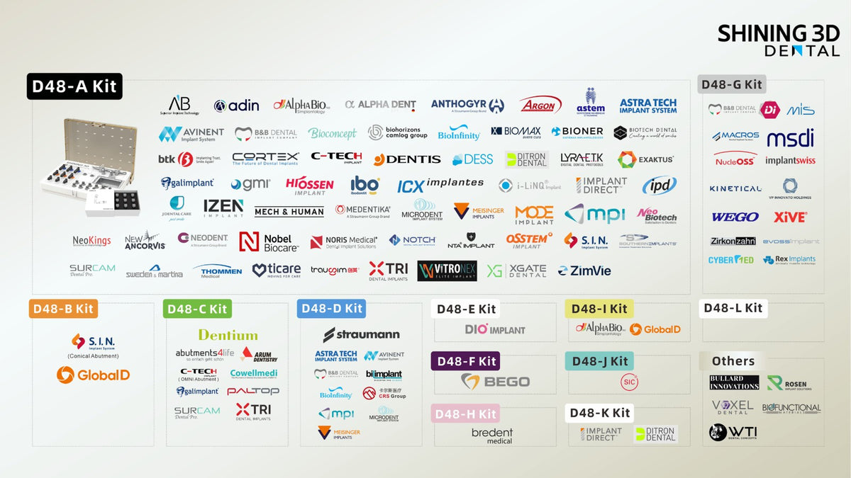
The high-accuracy coded scanbodies connect to compatible multi-unit abutments (MUA) and are specific to Elite and Elite Wireless. Confirm the implant library and physical interface before use. The open system exports data for compatible CAD software.